Limmer Education

by Chris Ebright
Our articles are read by an automated voice. We offer the option to listen to our articles as soon as they are published to enhance accessibility. Issues? Please let us know using the contact form.
The previous edition of Back to the Basics discussed the differences between ventilation and respiration.
As long as those physiological processes are functional, pulmonary capillaries can suck up the available alveolar oxygen and inject carbon dioxide in the opposite direction. Now, that is all well and good, but what if blood never circulated out of the lungs? You guessed it…WE. STILL. DIE. Remember, cellular functions require continuous oxygen delivery and carbon dioxide elimination.
The dictionary defines perfusion as “the passage of fluid through the lymphatic system or blood vessels to an organ or a tissue.” Fine, but for us EMS types, let’s make this a little more tangible: “Air goes in and out, blood goes round and round, any variation on this is bad.” Perfusion is the necessary component to transport the goods of ventilation and external respiration throughout the body. Take CPR, for example. You can ventilate the lungs with as much air as they can hold, but if you’re not compressing the chest and circulating nutrients and oxygen, the patient’s brain and tissues still deteriorate.
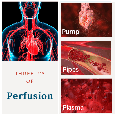
Perfusion is composed of what I call the Three P’s: The Pump (heart), the Pipes (blood vessels), and the Plasma (blood). Normal perfusion moves blood to the cellular capillary beds, where internal respiration of nutrients and oxygen then takes place. After passing through the cell membrane, oxygen binds onto the electron transport chain (ETS) within the mitochondria. Glucose (the best energy source) has already been broken down through a series of steps during glycolysis and the Krebs cycle (the medics reading that just had a small seizure). The products of the Krebs cycle, NADH and FADH2 pass their electrons through the ETS, where on the final protein complex, oxygen locks on – producing water, carbon dioxide, and finalizes ATP synthesis.

Each glucose molecule with oxygen present (aerobic metabolism) produces 32-34 ATP, providing energy to maintain cellular functions. Carbon dioxide diffuses from within the cell into the surrounding capillaries for its trip back to the lungs. It circulates through the venous system, and eventually into the pulmonary capillaries. However, when perfusion deteriorates and oxygen is not present (anaerobic metabolism), only two ATP result. The resultant lack of energy compromises all cellular functions.
Additionally, metabolic (lactic) acidosis, potassium, calcium and sodium imbalances, cellular membrane damage, intracellular and intravascular fluid shifts, hypercarbia, and hypercoagulation within the surrounding capillaries commences. Preventing this disaster takes all three “P’s” running efficiently all day, every day. Here is how they do it.
The average adult heart contracts between 60 and 100 times per minute. The stroke volume of each ventricular contraction averages 70 milliliters. Stroke volume is determined by preload (how much blood is available to fill the ventricle before contraction) and myocardial contractility. By multiplying the heart rate and stroke volume, the result is the amount of blood circulated per minute. This is cardiac output, which for the average adult is five to six liters per minute – supporting cellular oxygen delivery and carbon dioxide removal. Heart malfunction resulting from decreased contractility/myocardial infarcts, chest trauma, hypertension, arrhythmias, infection, or congenital defects, compromises adequate perfusion. Fixing this problem requires the EMT to address the specific heart issue(s) as best as possible, seeking advanced care when necessary.
Normal blood pressure equals good perfusion (even though there are numerous debates as to what “normal” is). Determining blood pressure is trickier, in the sense that you take the average cardiac output and multiply that value to the afterload. Put in simplest terms, afterload, also known as systemic vascular resistance (SVR), is the resistance the heart must overcome during a ventricular contraction to push blood into the systemic circulation. Blood vessel length, blood vessel diameter, and fluid (blood) viscosity all factor into its value. Vessel length is constant at the adult stage, so in this context, let’s discuss the other two.
As a vessel’s diameter decreases, resistance (afterload) increases. Resistance is highest in the capillary beds and lowest in the aorta. If a vessel excessively narrows, as in a chronically-hypertensive or pre-eclampsia patient, inadequate distal perfusion results. If the vessel does the opposite and over-dilates, as in spinal shock or anaphylaxis, afterload decreases, compromising blood pressure and systemic perfusion. The human body requires blood vessels to dilate and contract daily to maintain adequate perfusion. As you stand up, sit down, exercise, sleep, etc., various changes in body position and metabolism require vessels to adjust their diameter (also known as vascular tone). Like watchtower guards overlooking a prison yard, chemoreceptors and baroreceptors regularly monitor for perfusion changes, responding to “keep the peace.”
Chemoreceptors are located in the carotid arteries, aortic arch, and medulla oblongata. When blood oxygen and pH levels decrease, and carbon dioxide levels increase, these receptors activate. Their nerve impulses shut down parasympathetic nerves while at the same time increase the stimulation of sympathetic nerves. The result is increased heart rate and myocardial contraction force, equaling a rise in cardiac output. The same sympathetic stimulation also stimulates peripheral blood vessels to constrict, increasing afterload and preload. The result is improved perfusion, oxygen delivery, and carbon dioxide elimination.
The primary baroreceptors are located in the carotid sinus and aortic arch. Unlike chemoreceptors, though, they dilate or constrict blood vessels based upon blood pressure changes. If blood pressure rises above normal, baroreceptors stimulate a shutdown of the vasomotor centers (blood vessels dilate) and activate the cardiac inhibition centers (cardiac output decreases). Once the blood pressure declines back to normal range, the baroreceptors cease transmission. Adversely, if the blood pressure drops below normal, baroreceptors shut off, causing the vasomotor centers to activate (blood vessels constrict) and the cardiac acceleration centers to activate (cardiac output increases).
Viscosity of the blood is the other variable that determines afterload. Perfusion deteriorates as viscosity increases. In other words, the more concentrated the blood, the higher the resistance, and the slower the flow. Think of it this way: You have a cup containing plain water and another cup containing a milkshake. When you suck through a straw, it is much easier to drink the water than the milkshake because of the concentration difference. The same holds true concerning the blood. COPD patients with excessive amounts of red blood cells, patients with leukemia and lymphomas (excessive white cells), dehydrated patients, abnormal blood clotting, and hypothermia all cause the blood to “thicken.”
Something else of note: Blood belongs only in one place, and that is within the pipes, but pipes bust and patients bleed. If the pipes aren’t busted, they can excessively leak plasma into surrounding tissues. Septic and burn patients are notorious for this, typically requiring support with vasopressors. Squeezing the pipes, along with fluid support, reduces the size of leaky capillaries and helps restore adequate perfusion.
Just like the “leaky” patient, when a patient is bleeding internally, externally, or both, the result can be fatal. Decreased circulating blood volume reduces preload, which is the amount of volume returning to the right atrium. Lower preload diminishes stroke volume and cardiac output. Declining cardiac output reduces blood pressure and overall systemic perfusion, resulting in the series of cellular events discussed earlier. The vast majority of patients require only direct pressure and perhaps a tourniquet to control and stop external bleeding. Easy enough, but if bleeding is internal, there is not much EMS professionals can do except R and R (recognize and run). These are surgical cases and need to be transported to the closest, appropriate facility. Even though advanced providers can infuse IV normal saline, lactated ringers, blood, or plasma to restore preload, altogether, these measures may not be enough to restore adequate perfusion.
Now, let’s walk away from the acute, isolated events that cause a patient to bleed and focus on everyday living. Just by being alive, our metabolism and bodily functions deplete intracellular and intravascular volume. The easiest way for the body to restore volume and maintain perfusion is by external fluid intake, i.e., drinking. Ever wonder why you feel thirsty? When you sweat, for example, plasma volume is lost, and the blood thickens. The resultant slower flow is detected by the hypothalamus, which in turn performs two actions. First, it sets off the thirst mechanism, hopefully making you drink. Once you drink enough volume to replace what was lost, the hypothalamus shuts it down. Second, it releases antidiuretic hormone (ADH) from the posterior pituitary. ADH travels through the bloodstream and goes to work on the kidney tubules, where fluid retention/secretion takes place. More on that in a minute.
How is it, then, the average human can go three to seven days without any fluid intake? To answer that, we need to discuss a little physiology.
The major organ that helps regulate blood volume is the kidney, and it HATES to be under-perfused. So much, that it will use multiple hormones to restore adequate perfusion. This process is called the renin-angiotensin-aldosterone system (Yes, the medics are probably seizing at the thought of this – again). Whenever renal (kidney) perfusion pressure drops below normal, its ability to properly filter out toxins, among other things, is compromised - big time. This is the stimulus that causes the kidney to release erythropoietin (EPO) and renin into the bloodstream. EPO circulates throughout the bloodstream and targets bone marrow, where it stimulates the production of red blood cells. Blood volume is partially restored, but its concentration has thickened. Increasing plasma/water volume is the other requirement, which is fulfilled by renin.
Renin is a hormone with one job – find its cousin from the liver, angiotensinogen. When the family reunion takes place in the bloodstream, renin and angiotensinogen combine and form angiotensin 1. Once angiotensin 1 reaches the lungs, an enzyme (ACE) converts it into the hormone angiotensin 2. Angiotensin 2 leaves the lungs and circulates back to the kidneys. During its journey, the mere presence of angiotensin 2 causes vasoconstriction and enhancement of the thirst mechanism. Its major action, however, takes place within the adrenal cortex, where its presence releases another hormone called aldosterone.
Aldosterone leaves the adrenals, travels through the blood, down to the kidney tubules, and meets up with ADH. Remember, both of these hormones have ended up in the same place because of low renal perfusion, which likely resulted from poor systemic perfusion. In any sense, think of aldosterone as a vacuum and ADH as a key master. ADH circulates to a tubule, unlocks, then opens a closed “door” on the distal renal tubule, making it extremely permeable. Once the door is wide open, serum aldosterone sucks up as much sodium from the tubule as it can. The sodium shift from tubule into the bloodstream results in the endgame – water within the tubule follows sodium into the bloodstream and, given enough time, restores plasma volume to an adequate level.
The only downside is, it takes a long time to get from renin release to water retention. Like any hormone-based process, it is a series of multiple steps rather than one swift physiological reaction. Thus, if a patient is actively bleeding, severely dehydrated, or ailing from some other form of decompensated shock, the renin reaction is too slow to restore timely and adequate circulation. Other means, as discussed previously (fluids, etc.), need to be instituted for a positive patient outcome.
So that’s it. I hope this discussion makes perfusion a little more understandable. There is much more to it, but being able to assess for deficiencies of a patient’s Pump, Pipe, or Plasma component will go miles to aid you in rendering the best care possible. Assess, reassess, and adjust. Patients with perfusion issues rarely “stay in one place” for very long.
“perfusion.” com. 2020. https://www.dictionary.com/browse/perfusion May, 2020)
Spector, D. (March 8, 2018). Here’s how many days a person can survive without water. Business Insider. Retrieved from: https://www.businessinsider.com/how-many-days-can-you-survive-without-water-2014-5

Limmer Education

Dan Limmer, BS, NRP

Limmer Education
Excellent - down to earth explanation, thank you